California by Optos®

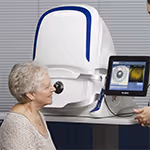
BBR Optometry are proud to offer the very latest in retinal imaging technology, with the Optos® California:

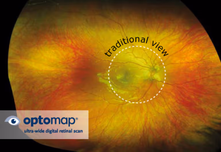
The Optos® California scans your eye to produce a digital image of the retina, called an optomap®.
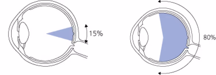
One quick scan shows 80% of a retina, helping your Optometrist to immediately identify potential health issues

A drawing of a pair of glasses with a blue light coming out of itA close up of a person's eye with the words traditional view on it

Without Optos®
Traditional scans can only record about
15% of the retina at any one time

The ultra-widefield Optos® scan

The optomap® shows 80% of a retina

The ultra-wide field of view considerably speeds up the screening process, helping your Optometrist to quickly identify potential issues and recommend a course of action that will minimise future problems.

Protect your vision and your health with an Optos® scan at BBR Optometry

CALIFORNIA By Optos®

What it does

  • Ultrawide digital image
  • Optomaps

How it works

  • Wide angle parabolic mirror
  • 2 lasers (deep red and green)

Benefits

  • 5x more retina visible than other imaging
  • 200° retinal image
  • Reference to compare in future
  • Looks beneath the surface
  • Rarely needs dilating drops
  • Assesses how cells work as well

Rules out

  • Macular degeneration
  • Diabetes
  • Retinal breaks, tears and detachments

If detected early, most retinal conditions and other diseases can be treated successfully.

Triton and Maestro 3D OCT

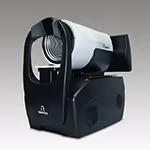
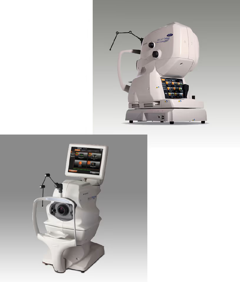
Optical Coherence Tomography (OCT) is the next generation in imaging the internal structures of the eye. The unique way the image is formed means that for the first time we can actually see below the surface of the retina and view the microscopic layers beneath. This is of profound importance in determining the precise diagnosis of visual problems and in helping us guide patients on the treatment options possible.

Once again BBR Optometry are able to offer its patients direct access to the most sophisticated eye examination available.

3D OCT imaging is automatically part of your BBR private Eye Examination and is included in our extension to the NHS Sight Test.

Typical conditions requiring OCT:

  • Macular degeneration or drusen
  • Macular hole or epiretinal membrane
  • At risk glaucoma
  • Vitreous Floaters
  • Diabetic retinopathy
  • Optic nerve disease

What it does

  • Optical Coherence Tomography
  • Digital 3D image

How it works

  • 256 scans in less than 5 seconds
  • Flash photo-3D image

Benefits

  • Shows detail that lies beneath
  • Incredible precision 1/10000mm
  • Things previously invisible
  • Nothing takes us by surprise
  • All previous images viewable

Rules out

  • Macular degeneration
  • Glaucoma detection and predict
Triton and Maestro eye test

BBR Optometry was the first optometry practice to bring retinal imaging to the Hereford high street (Polaroid film 1989) which became digital in 2000.

Henson

Henson by Elektron

What it does

  • Maps side vision
  • LED lights different brightness

How it works

  • Easier operator controlled system, just count

Benefits

  • You don’t notice loss of side vision
  • Fast screening
  • Vital in glaucoma detection & monitoring

Rules out

  • Glaucoma risk
  • Progressive change

Humphrey by Carl Zeiss Meditec

What it does

  • Maps side vision
  • Projected spots of light

How it works

  • You control speed of response

Benefits

  • You don’t notice loss of side vision
  • Fast screening
  • Vital in glaucoma detection & monitoring

Rules out

  • Glaucoma risk
  • Progressive change
humphrey
i care eye test equipement

i-Care by I-care Finland Oy

What it does

  • Measures fluid pressure in eye

How it works

  • Tiny probe just kisses the eye surface many times
  • Internal averaging system eliminates false readings

Benefits

  • No puff of air
  • Perfect sterility
  • No drops
  • Can keep soft lenses in

Important for

  • Risk factors for glaucoma

Aladdin by Topcon

What it does

  • Maps shape of front of the eye
  • Measures the length of the eye ball
  • Determines the depth and thickness of eye structures

How it works

  • Reflects circular rings of light onto the surface of the eye
  • Uses light interreference to take depth and thickness measurements
  • Computer technology analyses data to create report

Benefits

  • Used for specialist contact lens fitting
  • Monitors cornea (front of the eye) shape
  • Monitors eye length growth for myopia (short-sightedness) in children

Important for

  • Conditions affecting corneal shape
topographer
Dry Eye Scanner

DNEye scanner by Rodenstock/Visionix

What it does

  • Measures shape of front of eye
  • Measures visual image quality for glare

How it works

  • Measures the transmission and reflectance of laser light through the eye
  • In normal and low light conditions

Benefits

  • No contact with eye
  • Does not need dilation of pupils

Important for

  • Best possible spectacle vision

Pachymeter by Pachmate DGH Tech

What it does

  • Measurement of thickness of membrane at front of eye

How it works

  • 1000’s of ultrasound scans
  • Measures thickness to 1/10000mm

Benefits

  • Essential to calculate glaucoma risk
  • Without it pressure measurements are guesswork

Important for

  • True high eye pressure from false
pachymeter
A close up of a pair of binoculars

Topcon’s CV-5000PRO

What it does

  • Replaces the trial frame on your nose
  • Enables remote control lens changes
  • Is easy to keep clean

How it works

  • All the lenses are stored behind sealed units
  • Controlled by optometrist using computer
  • Electric motors change lenses in less than 1/100th second

Benefits

  • Easier to keep clean
  • Not so heavy on your nose
  • Easier to show the "before" and "after" test results

Important for

  • Minimises contamination risk
  • Difficult to clean loose lenses
  • Easier to decide if vision is better